
Researchers Assess Gold Standard for Peyronie’s Disease Measurement for Repeatability
Background
Peyronie’s disease (PD) is a connective tissue disorder that involves the buildup of fibrotic plaque in the penis, which can lead to penile curvature, deformities like indentations or an hourglass shape, and a loss of elasticity. As a result, many men with PD experience painful erections or erectile dysfunction (ED).
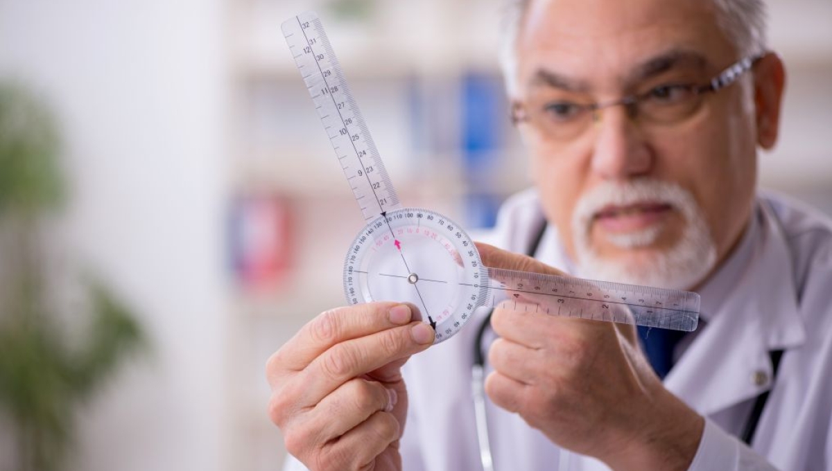
The current gold standard for assessing PD curvature involves in-office intracavernous injections (ICI) to induce an erection and measure the degree of curvature with a goniometer. However, the recommendations provided by the American Urological Association (AUA), Canadian Urological Association (CUA), and European Urological Association (EUA) do not provide strict guidelines on how to measure curvature in practice, which can lead to inconsistency among clinicians.
Study Objectives
- To assess the reproducibility of curvature measurements across common measurement methods.
- To highlight the need for more detailed standardization to enhance comparability in clinical care and research.
Methods
- The same 22 men submitted two sets of images of their erect penis, with each set including both the dorsal and lateral angles.
- One set of images was taken at home by the patient with specific instructions.
- Use of a constriction band was required.
- Images were taken against a provided sheet of laminated graph paper.
- One set of images was taken at the clinic following the standard ICI-induced erection, also using a constriction band and graph paper.
- Doppler ultrasound examined penile tissues to ensure rigidity following ICI.
- Images were printed and anonymized.
- Four urologists with an average of 16.25 years of experience diagnosing and treating PD were provided with different sets of images.
- Urologists 1 and 2 were to measure the patient-provided images without assisting lines (Kelami method).
- Urologists 3 and 4 were to measure the clinic-provided images without assisting lines.
- 2-4 weeks later, the images were measured by the opposite group of urologists, this time using assisting lines.
- Urologists 1 and 2 measured the clinic-provided images.
- Urologists 3 and 4 measured the patient-provided images.
- Each round of measuring included 3 separate measurements from each urologist, which were averaged before analysis.
- Researchers used an interclass correlation coefficient (ICC) and scatterplots to assess repeatability.
- One set of images was taken at home by the patient with specific instructions.
Key Results/Analysis
- Averages for both dorsal and lateral measurements were higher for the clinic-provided images than for the patient-provided images (50.16°-61.22° vs 41.47°-47.17° lateral, 27.36°-33.17° vs 25.38°-26.48° dorsal).
- This may have been due to different interpretations of instructions given to patients, or less reliable rigidity from patient-induced erections compared with ICI-induced erections.
- The scatterplots were relatively consistent, and ICCs were relatively high across all measurement types except for the clinic-provided image assessment using assisting lines.
- Scatterplots revealed a linear correlation for patient-provided images with and without lines, and clinic-provided images without lines.
- Comparing urologists 1 and 2, there appeared to be only moderate repeatability.
- This means PD assessors may have a more difficult time assessing curvature using images taken in-clinic with ICI-induced erections.
- Researchers question the Kelami method for assessing PD using assisting lines, saying it may not be entirely helpful anymore, despite influential principles.
- Researchers found there is still no consistent advantage of at-home over in-clinic assessments for initial PD inquiries.
- At-home options may be beneficial for patients who are apprehensive of ICI, because they still provide reasonable consistency in measurement.
- Variability among the assessors shows that images alone are not enough to replace in-office assessments completely.
- Previous research has shown similar concerns about the repeatability of PD assessments, which is consistent with the lack of standardization in AUA, CUA, and EUA guidelines. A “second opinion” may not be entirely accurate for the patient if the assessment systems differ widely between clinicians.
- Researchers believe the results of this study may provide areas for further research, such as:
- Assessing best practices for the standardization of PD curvature measurement.
- Testing the reliability of technological advancements like app-based or AI-assisted PD assessments.
- Looking into the reliability of the same assessor over time.
Key Takeaways
- Patient-provided images can still be useful for initial PD assessments but should not be considered the gold standard.
- There is still plenty of room for improvement when it comes to PD assessment and standardization of measurement processes.
- Future studies should look into technology-assisted assessment programs and further standardization of guidelines.
Resources
Wiborg, M. H., Krøijer, R., Kallestrup, E. B., Laursen, B. S., Berg-Beckhoff, G., & Lund, L. (2025). Repeatability in measuring curvature in Peyronie’s disease. Sexual Medicine, 13(6). https://doi.org/10.1093/sexmed/qfaf105
Public
Providers
Members Only
Newsletter
ISSM Update
Sign up here to receive ISSM Updates and stay informed about sexual medicine, ISSM news, events, and more.